Murali K
Tagline:Faculty at IIT Madras | Exploring light based imaging for cancer detection and brain health

About the PI
Dr. Murali completed his Ph.D. in Biomedical Engineering at the Indian Institute of Technology (IIT) Bombay. He subsequently pursued postdoctoral training at the Athinoula A. Martinos Center for Biomedical Imaging, Department of Radiology, Massachusetts General Hospital, and later at the Department of Otolaryngology–Head and Neck Surgery, Massachusetts Eye and Ear, Harvard Medical School. He is currently an Assistant Professor in the Department of Medical Sciences and Technology, IIT Madras.
Before transitioning to research, he was part of Medtronic India, providing clinical support and training to operating room (OR) staff on advanced endo-mechanical stapling devices and related OR technologies.
His research lies at the intersection of biomedical optics, system development, and algorithm design, aiming to advance optical imaging for applications in oncology and neuroimaging. Key research areas include tumor visualization and blood flow imaging, with an emphasis on non-invasive approaches to quantify clinically relevant biomarkers.
About the lab
Students joining the group will have the opportunity to pursue projects across optics, computation, and clinical translation. Depending on their background and interests, this may include the design of optical imaging systems, development of clinical and preclinical imaging protocols, implementation of tomographic reconstruction algorithms, hands-on preclinical studies involving procedures such as surgery and blood-flow monitoring, and participation in clinical collaborations for image-guided surgery and diagnostics.
Passionate about biomedical optics and imaging?
Reach out with your CV and a short note on your interests. We welcome enthusiastic students and researchers to join our growing team. Please contact the PI via email.
MS (by Research) and PhD Admissions (2026–27)
Applications are now open for the M.S. (by Research) and PhD programmes at the Indian Institute of Technology Madras.
Prospective students interested in joining the lab should apply through the IIT Madras Research Admissions Portal.
📅 Admission Cycles
July 2026 (Fall cycle): Application deadline 30 March 2026
January 2027 (Spring cycle): Applications open 31 March 2026 – 30 October 2026
Applicants may apply to M.S. (by Research) or PhD programmes, including Direct PhD after a Bachelor’s degree.
For eligibility criteria and application details, please refer to the department website:
🔗 https://mst.iitm.ac.in/ms-phd-applications/
Apply through the IIT Madras Research Admissions Portal:
🔗 https://research.iitm.ac.in
For any questions related to working with the group, please write to me by email.
ANRF National Postdoctoral Fellowship (NPDF) — Call for Applications (2026)
The Call for Applications for the ANRF National Postdoctoral Fellowship Scheme (NPDF) will be open from 15 January 2026 to 17 February 2026.
Postdoctoral researchers interested in working with my group under the NPDF scheme are invited to get in touch. The fellowship supports independent research aligned with the group’s focus areas.
Applicants are encouraged to review the detailed fellowship guidelines and eligibility criteria at:
https://anrfonline.in/ANRF/npdf
Interested candidates may email k_murali@iitm.ac.in to discuss project ideas and the application process.
Summer Internship Opportunities (IIT Madras Summer Fellowship Programme)
Interns at IIT Madras are recruited through the Summer Fellowship Programme (SFP). Interested candidates must apply via the official portal.
Important Note: Students from IITs are not eligible to apply.
Application Deadline: 02 March 2026, 5:00 PM
Eligibility Criteria:
Candidates pursuing 3rd year of UG (BE/ B.Tech / B.Sc Engg./3-year B.Sc. with Chemistry, Physics & Mathematics) or 3rd or 4th year of Integrated / Dual Degree in Masters Programme (M.E./ M.Tech / M.Sc) 1st year of M.Tech/ M.A,/ MBA with outstanding academic background in terms of high ranks in University examinations are encouraged to apply, highlighting their academic performance and achievement including papers presented at seminars, projects executed, design contests participated, score/rank in Mathematics Olympiad and any other awards/distinctions obtained.
Period of the Summer Fellowship Programme: The programme is likely to commence from 18th May 2026 to 17th July 2026.
Stipend: A sum of Rs.15000/- per month will be given as stipend for a maximum period of 2 months, prorated for the actual period of internship at IIT Madras.
Hostel: Hostel and Mess facility will be provided on payment basis, subject to availability.
Application Link:
Selected Journal Publications
High-Speed Wide-Field Fluorescence Lifetime Imaging for Intraoperative Tumor Visualization and In Vivo Multiplexing
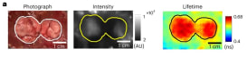
Journal ArticlePublisher:Biomedical Optics ExpressDate:2025Authors:Murali KrishnamoorthyRahul PalSteven HouHak Soo ChoiBrian J BacskaiAlexei A Bogdanov JrKenneth K TanabeMark A VarvaresAnand TN KumarDescription:We present a technique for fast wide-field fluorescence lifetime (FLT) imaging using simultaneous steady-state and time- (or frequency-) domain measurements acquired in a single shot and exploiting the theoretical dependence of fluorescence intensity on FLT. Using theory, simulations, and in vitro experiments, we show that the single-shot FLT (S-FLT) method can provide absolute FLTs at a higher signal-to-noise ratio compared to other high-speed FLT imaging techniques, without the need for sample-dependent system calibration or model training. We demonstrate that S-FLT can provide accurate tumor delineation in real-time during surgical resections in preclinical tumor models in vivo and in clinical patient specimens ex vivo. We also extend S-FLT for real-time multiplexing and demonstrate this technique for quantifying dynamic FLT changes in vitro and for multiplexing of organ-targeted NIR fluorophores in vivo. Our results suggest that S-FLT can enable real-time quantitative high-throughput studies in preclinical applications and accurate tumor margin delineation in real-time during surgeries.
Fluorescence Lifetime Imaging Enables In Vivo Quantification of PD-L1 Expression and Intertumoral Heterogeneity
Journal ArticlePublisher:Cancer ResearchDate:2025Authors:Rahul PalMurali KAya MatsuiHoman KangSatoru MoritaHajime TaniguchiTatsuya KobayashiAtsuyo MoritaHak Soo ChoiDan G DudaothersDescription:Patient selection for cancer immunotherapy requires precise, quantitative readouts of biomarker expression in intact tumors that can be reliably compared across multiple subjects over time. The current clinical standard biomarker for assessing immunotherapy response is PD-L1 expression, typically quantified using IHC. This method, however, only provides snapshots of PD-L1 expression status in microscopic regions of ex vivo specimens. Although various targeted probes have been investigated for in vivo imaging of PD-L1, nonspecific probe accumulation within the tumor microenvironment has hindered accurate quantification, limiting the utility for preclinical and clinical studies. Here, we demonstrated that in vivo time-domain fluorescence imaging of an anti–PD-L1 antibody tagged with the near-infrared fluorophore IRDye 800CW (αPDL1-800) can yield quantitative estimates of baseline tumor PD-L1 heterogeneity across untreated mice, as well as variations in PD-L1 expression in mice undergoing clinically relevant anti–PD-1 treatment. The fluorescence lifetime (FLT) of PD-L1–bound αPDL1-800 was significantly longer than the FLT of nonspecifically accumulated αPDL1-800 in the tumor microenvironment. This FLT contrast allowed quantification of PD-L1 expression across mice both in superficial breast tumors using planar FLT imaging and in deep-seated liver tumors (>5 mm depth) using the asymptotic time-domain algorithm for fluorescence tomography. These findings suggest that FLT imaging can accelerate the preclinical investigation and clinical translation of new immunotherapy treatments by enabling robust quantification of receptor expression across subjects.
Fluorescence lifetime of injected indocyanine green as a universal marker of solid tumours in patients
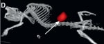
Journal ArticlePublisher:Nature Biomedical EngineeringDate:2023Authors:Rahul PalThinzar M LwinMurali KHannah R CollinsCorey D ChanAndrey PrilutskiyMacLean P NasrallahTom H DijkhuisShriya ShuklaAmy L KendallothersDescription:The surgical resection of solid tumours can be enhanced by fluorescence-guided imaging. However, variable tumour uptake and incomplete clearance of fluorescent dyes reduces the accuracy of distinguishing tumour from normal tissue via conventional fluorescence intensity-based imaging. Here we show that, after systemic injection of the near-infrared dye indocyanine green in patients with various types of solid tumour, the fluorescence lifetime (FLT) of tumour tissue is longer than the FLT of non-cancerous tissue. This tumour-specific shift in FLT can be used to distinguish tumours from normal tissue with an accuracy of over 97% across tumour types, and can be visualized at the cellular level using microscopy and in larger specimens through wide-field imaging. Unlike fluorescence intensity, which depends on imaging-system parameters, tissue depth and the amount of dye taken up by tumours, FLT is a photophysical property that is largely independent of these factors. FLT imaging with indocyanine green may improve the accuracy of cancer surgeries.

Spatio-temporal Simulation of Laser Speckles using Stochastic Differential Equations
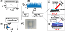
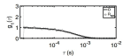
Journal ArticlePublisher:IEEE Journal of Selected Topics in Quantum ElectronicsDate:2025Authors:Soumyajit SarkarK MuraliAnindita ChandraSusweta DasHari M VarmaDescription:Laser speckle imaging modalities are receiving considerable attention for applications in both superficial and deep tissue imaging. This growing interest arises from their straightforward and cost-effective instrumentation, as well as the ease of computational implementation. Recent developments in this field require the calibration of devices, which often necessitates the simulation of dynamic laser speckles in tissue. Previous simulation models have predominantly relied on statistical tools such as copulas or Fourier transform methods based on diffraction theory. In our recent work, we introduced the use of Stochastic Differential Equations (SDEs) to model the temporal evolution of speckles, utilizing a predetermined probability density function and temporal autocorrelation. We further extended this simulation to create a calibrating phantom by inputting the stochastic time series data generated by SDEs into a piezo actuator. In the previous studies, the time evolution of each pixel was modeled independently using multiple SDEs. In the current work, we introduce a method for generating spatio-temporal dynamics of speckles characterized by predefined spatial and temporal correlation structures. This approach integrates SDE with Cholesky decomposition and inverse sampling method to achieve the desired spatial correlation profiles. The simulation results are obtained for practically feasible experimental parameters, effectively generating speckles that closely approximate the values observed in experimental settings.
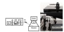
Optimized laser speckle–based imaging system and methods for deep tissue cerebral blood flow imaging in small rodents
Journal ArticlePublisher:NeurophotonicsDate:2025Authors:Ria PaulSoumyajit SarkarSusweta DasShruti D MaratheMurali KNixon M AbrahamHari M VarmaDescription:Significance: The imaging of cerebral blood flow in small rodents is crucial for a better understanding of brain functions in healthy and diseased conditions. Existing methods often struggle to provide both superficial and deep tissue blood flow measurements in a non-invasive, flexible, and reliable manner, creating a need for an integrated platform that addresses these limitations.
Aim: We aim to design and develop a multi-modal laser speckle–based imaging platform and associated algorithms to image superficial and deep tissue cerebral blood flow in small rodents.
Approach: A modular design has been adopted to integrate laser speckle contrast imaging and multi-speckle diffuse correlation tomography to a single cerebral blood flow imaging platform for small rodents with an independent module for animal holding and handling. A topographic imaging method, equipped with a filter to remove surface artifacts, was incorporated to image cerebral blood flow changes in response to forepaw and olfactory stimuli activations, with the skull and scalp kept intact.
Results: A significant increase in blood flow was found in the olfactory bulbs of mice post-stimulation by various odors (p<0.01). Similarly, forepaw stimulation resulted in a significant increase in blood flow in the contralateral side of the somatosensory cortex with the application of the filter for skull and scalp intact, skull intact, and skull removed cases (p<0.01).
Conclusions: We have validated our system through functional studies, demonstrating its capability to detect enhanced blood flow changes across the olfactory bulbs and somatosensory cortex in rodents with potential for broad applications in preclinical research.
Miniaturized Fab’imaging probe derived from a clinical antibody: characterization and imaging in CRISPRi-attenuated mammary tumor models
Journal ArticlePublisher:iScienceDate:2024Authors:Suresh GuptaRahul PalEric J SchmidtMurali KrishnamoorthyAnita LeporatiAnand TN KumarAlexei BogdanovDescription:Highlights:
• Both fluorescence intensity and lifetime increase after fragmentation of the Fab’ NIR probe
• Fluorescence lifetime contrast increases upon internalization of Fab’ NIR probe
• CRISPRi yielded two times lower EGFR expression in triple-negative breast cancer cells
• Fab’ probe showed the feasibility of CRISPRi effect imaging in vivo at an early time pointSummary
Clinical imaging-assisted oncosurgical navigation requires cancer-specific miniaturized optical imaging probes. We report a near-infrared (NIR) Fab’-based epidermal growth factor receptor (EGFR)-specific probe carrying 3 NIR fluorophores (Fab’-800CW), which retained high-affinity binding to EGFR ectodomain (equilibrium KDE = 1 nM). Fab’-800CW showed a robust 4-times gain of fluorescence intensity (FI) and a 20% lifetime (FLT) increase under the conditions mimicking intracellular degradation. The probe was tested by using triple-negative breast cancer (TNBC) cell lines obtained by applying CRISPR interference (CRISPRi) effect of EGFR-targeting sgRNA and dCas9-KRAB chimera coexpression in MDA-MB-231 cells (WT cells). FI imaging in cell culture proved a 50% EGFR expression attenuation by CRISPRi. FI imaging in animals harboring attenuated or WT TNBC tumors with ex vivo corroboration identified differences between WT and CRISPRi tumors FI at 30 min post injection. Our results suggest the feasibility of EGFR expression imaging using a Fab’-based probe relevant for imaging-guided cancer surgery.Tunable dynamical tissue phantom for laser speckle imaging
Journal ArticlePublisher:Biomedical Optics ExpressDate:2024Authors:Soumyajit SarkarMurali KHari M VarmaDescription:We introduce a novel method to design and implement a tunable dynamical tissue phantom for laser speckle-based in-vivo blood flow imaging. This approach relies on stochastic differential equations (SDE) to control a piezoelectric actuator which, upon illuminated with a laser source, generates speckles of pre-defined probability density function and auto-correlation. The validation experiments show that the phantom can generate dynamic speckles that closely replicate both surfaces as well as deep tissue blood flow for a reasonably wide range and accuracy.
Laser speckle simulation tool based on stochastic differential equations for bio imaging applications
Journal ArticlePublisher:Biomedical Optics ExpressDate:2022Authors:K MuraliHari M VarmaDescription:Laser speckle-based blood flow imaging is a well-accepted and widely used method for pre-clinical and clinical applications. Although it was introduced as a method to measure only superficial blood flow (< 1mm depth), several recently introduced variants resulted in measuring deep tissue blood flow (a few cm) as well. A means of simulating laser speckles is often necessary for the analysis and development of these imaging modalities, as evident from many such attempts towards developing simulation tools in the past. Such methods often employ Fourier transforms or statistical tools to simulate speckles with desired statistical properties. We present the first method to use a stochastic differential equation to generate laser speckles with a pre-determined probability density function and a temporal auto-correlation. The method allows the choice of apriori gamma distribution along with simple exponential or more complex temporal auto-correlation statistics for simulated speckles, making it suitable for different blood flow profiles. In contrast to the existing methods that often generate speckles associated with superficial flow, we simulate both superficial and diffuse speckles leading to applications in deep tissue blood flow imaging. In addition, we have also incorporated appropriate models for noise associated with the detectors to simulate realistic speckles. We have validated our model by comparing the simulated speckles with those obtained from in-vivo studies in mice and healthy human subject.
High-density diffuse correlation tomography with enhanced depth localization and minimal surface artefacts
Journal ArticlePublisher:Biomedical Optics ExpressDate:2022Authors:Ria PaulK MuraliHari M VarmaDescription:A spatially weighted filter applied to both the measurement and the Jacobian is proposed for high-density diffuse correlation tomography (DCT) to remove unwanted extracerebral interferences and artefacts along with better depth localization in the reconstructed blood flow images. High-density DCT is implemented by appropriate modification of recently introduced Multi-speckle Diffuse Correlation Spectroscopy (M-DCS) system. Additionally, we have used autocorrelation measurements at multiple delay-times in an iterative manner to improve the reconstruction results. The proposed scheme has been validated by simulations, phantom experiments and in-vivo human experiments.
A simple algorithm for diffuse optical tomography without Jacobian inversion
Journal ArticlePublisher:Biomedical Physics & Engineering ExpressDate:2022Authors:Ria PaulK MuraliSumana ChetiaHari M VarmaDescription:A computationally simpler algorithm to reconstruct the optical property distribution of turbid media using diffuse optical tomographic principles is presented. The proposed algorithm eliminates the requirement of large Jacobian matrix inversion which otherwise is essential for tomographic imaging. The most significant Jacobians are identified based on proper thresholding of the measurement and the intersection of these Jacobians gives the approximate spatial location of the inhomogeneity. The algorithm is tested and optimized using simulations and further validated using tissue-mimicking phantom-based experiments and in-vivo small-animal experiments.
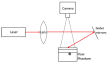
Multi-speckle diffuse correlation spectroscopy to measure cerebral blood flow
Journal ArticlePublisher:Biomedical Optics ExpressDate:2020Authors:K MuraliHari M VarmaDescription:We present a multi-speckle diffuse correlation spectroscopy (DCS) system for measuring cerebral blood flow in the healthy adult human brain. In contrast to the need for a high frame rate camera to measure the multi-speckle intensity auto-correlation, we employ a low frame rate camera to measure the auto-correlation using the recently introduced multi-step volterra integral method (MVIM). The results are validated by comparison against the blood flow measured using standard DCS system.
On the equivalence of speckle contrast-based and diffuse correlation spectroscopy methods in measuring in vivo blood flow
Journal ArticlePublisher:Optics LettersDate:2020Authors:K MuraliAK NandakumaranHari M VarmaDescription:We establish the equivalence between laser speckle contrast-based and diffuse correlation spectroscopy methods in in vivo imaging of blood flow using the Volterra integral equation theory. We further substantiate the need of regularized fitting while employing the multiexposure speckle contrast imaging to recover autocorrelation function.
Recovery of the diffuse correlation spectroscopy data-type from speckle contrast measurements: towards low-cost, deep-tissue blood flow measurements
Journal ArticlePublisher:Biomedical Optics ExpressDate:2019Authors:K MuraliAK NandakumaranTurgut DurduranHari M VarmaDescription:A multi-step Volterra integral equation-based algorithm was developed to measure the electric field auto-correlation function from multi-exposure speckle contrast data. This enabled us to derive an estimate of the full diffuse correlation spectroscopy data-type from a low-cost, camera-based system. This method is equally applicable for both single and multiple scattering field auto-correlation models. The feasibility of the system and method was verified using simulation studies, tissue mimicking phantoms and subsequently in in vivo experiments.


Contact
Address
Department of Medical Sciences & Technology
5th Floor, Room - 552, New Academic Complex (NAC)-II
Indian Institute of Technology Madras (IITM)
Chennai, Tamil Nadu-600036, India